

ABSTRACT
Background and Aim: Antimicrobial resistance (AMR) has emerged as a major threat to animal and public health. Escherichia coli is an important foodborne pathogen and a causative agent of subclinical mastitis in dairy animals. In Siliragung Subdistrict, Banyuwangi, Indonesia, fresh goat milk is commonly consumed, creating potential risks for the transmission of resistant bacteria. This study investigated the occurrence and AMR patterns of E. coli isolated from raw goat milk obtained from goats with subclinical mastitis.
Materials and Methods: A cross-sectional study was conducted between June and December 2024 involving 224 lactating goats from seven dairy goat farms in Siliragung Subdistrict, Banyuwangi, Indonesia. Subclinical mastitis was screened using the California Mastitis Test, and samples with scores of 3 and 4 were subjected to bacteriological examination. Presumptive E. coli isolates were identified using colony morphology, Gram staining, and indole, methyl red, Voges–Proskauer, and citrate biochemical tests. Antimicrobial susceptibility was evaluated by the Kirby–Bauer disk diffusion method against ampicillin, cephalothin, streptomycin, ciprofloxacin, erythromycin, chloramphenicol, trimethoprim, and tetracycline according to Clinical and Laboratory Standards Institute (CLSI) guidelines. Multidrug resistance (MDR) was defined as resistance to at least three antimicrobial classes.
Results: Among 224 raw goat milk samples, 91 (40.6%) were positive for subclinical mastitis. Thirty isolates (32.9%) were identified as E. coli. The highest resistance rates were observed for erythromycin (63.3%) and tetracycline (53.3%), whereas the highest susceptibilities were recorded for ciprofloxacin (76.7%) and trimethoprim (60.0%). Ten isolates (33.3%) exhibited MDR. Marked variation in MDR prevalence was observed among farms, ranging from 0% to 50%.
Conclusion: Raw goat milk from goats with subclinical mastitis in Banyuwangi harbored antimicrobial-resistant and multidrug-resistant E. coli. The predominance of erythromycin- and tetracycline-resistant bacteria and the substantial MDR rate emphasize the need for prudent antimicrobial use, improved farm biosecurity, routine susceptibility testing, and integrated One Health surveillance to reduce the risk of dissemination of resistant bacteria through the dairy goat production system.
Keywords: antimicrobial resistance, Banyuwangi, dairy goats, Escherichia coli, multidrug resistance, public health, raw milk, subclinical mastitis, zoonotic risk.
INTRODUCTION
Antimicrobial resistance (AMR) is a major challenge affecting both human and animal health worldwide [1]. In South Asia alone, AMR has been associated with approximately 389,000 deaths, annual livestock production losses of up to 7.5%, a projected 3.8% decline in global exports, and cumulative healthcare costs estimated to reach US$1 trillion by 2050 [2]. The inappropriate and excessive use of antibiotics promotes the emergence and dissemination of resistant bacteria. Antibiotics are commonly used to treat mastitis and other bacterial infections caused by Escherichia coli in humans and animals. Several antimicrobial agents are routinely used for this purpose, including ampicillin, cephalothin, streptomycin, ciprofloxacin, erythromycin, chloramphenicol, trimethoprim, and tetracycline [3, 4]. Mastitis remains one of the most important diseases affecting dairy production, including goat farming, and E. coli is recognized as a principal bacterial pathogen associated with the condition [5]. Mastitis causes substantial economic losses because of decreased milk production, discarded milk, and increased treatment costs. In particular, subclinical mastitis is frequently treated with various antibiotics, which may contribute to the development and spread of AMR among bacterial populations [6].
E. coli, a member of the family Enterobacteriaceae, has ruminants as one of its major reservoirs and can reach humans through the food chain [7]. This bacterium is among the leading causes of foodborne diseases worldwide, and milk and dairy products constitute important vehicles for its transmission to humans [8]. Exposure of E. coli to antibiotics in livestock environments may facilitate the development of resistance and the transfer of resistance genes to other E. coli strains [9]. Consequently, the emergence of antibiotic-resistant E. coli has become a major public health concern, with contaminated milk serving as an important source of resistant strains [10]. Antibiotic-resistant E. coli was estimated to cause approximately 200,000 deaths worldwide in 2019 [11]. Ranjbar et al. [12] reported a prevalence of 30.16% for antibiotic-resistant E. coli isolated from milk and dairy products. Commonly used broad-spectrum antibiotics include ampicillin, chloramphenicol, erythromycin, tetracycline, and trimethoprim [12]. In addition, streptomycin and ciprofloxacin are primarily active against Gram-negative bacteria, whereas cephalothin is effective against many Gram-positive bacteria and certain Gram-negative cocci, including E. coli [13, 14].
Consumption of fresh milk contaminated with antibiotic-resistant E. coli may facilitate the transfer of antibiotic resistance genes to the human gastrointestinal tract [15]. Milk and dairy products are valuable sources of proteins, lipids, minerals, and vitamins and therefore contribute significantly to human nutrition [16]. Moreover, milk represents an excellent medium for the growth of both pathogenic and nonpathogenic microorganisms [17]. Interest in goat milk and goat milk products has increased due to the growing demand for healthy foods and their suitability for individuals with allergies or intolerances to cow’s milk [18]. Although goat milk resembles cow milk in appearance and taste, it possesses distinct chemical, biochemical, physical, and nutritional characteristics [19]. One notable difference is the presence of smaller fat globules in goat milk, which facilitate more rapid digestion by digestive enzymes [20].
In Indonesia, antimicrobial-resistant E. coli has been reported in several animal-derived food products, including poultry meat and raw milk, indicating widespread antibiotic exposure of bacteria in livestock production systems [21]. In Banyuwangi Regency, antimicrobial-resistant bacteria have previously been detected in dairy goat farms, particularly Staphylococcus aureus isolated from cases of subclinical mastitis [22]. These findings indicate that AMR is already established within local dairy production systems. Siliragung Subdistrict, located in the southern region of Banyuwangi Regency, is characterized by the widespread consumption of fresh goat milk by the local community [23]. The area contains seven dairy goat farms raising local breeds such as Jawa Randu, Sapera, and Peranakan Etawa goats, with a total productive population of 224 animals [24].
Although previous studies have documented antimicrobial-resistant bacteria in dairy goat farming systems in Banyuwangi and reported the occurrence of E. coli in animal-derived food products in Indonesia [21, 22], information regarding the phenotypic AMR profiles of E. coli isolated specifically from raw goat milk associated with subclinical mastitis remains scarce. Earlier investigations were limited to other bacterial pathogens, mixed animal species, or broader geographical areas and did not provide farm-level resistance patterns for E. coli in dairy goats. Moreover, no previous study has comprehensively evaluated the susceptibility of E. coli isolates obtained from all active dairy goat farms in Siliragung Subdistrict, an area where fresh goat milk is traditionally consumed and where the risk of transmission of resistant bacteria to humans may be substantial. Therefore, detailed local data are needed to understand the occurrence and resistance characteristics of E. coli in this production system and to support evidence-based antimicrobial stewardship strategies within a One Health framework.
Therefore, this study aimed to investigate the occurrence and AMR patterns of E. coli isolated from raw goat milk obtained from goats with subclinical mastitis in Siliragung Subdistrict, Banyuwangi Regency, Indonesia. Specifically, the study evaluated the prevalence of E. coli among milk samples collected from seven dairy goat farms and determined the susceptibility profiles of the isolates against commonly used antimicrobial agents. Furthermore, the study assessed the occurrence of MDR isolates to provide baseline data for local surveillance and to support prudent antimicrobial use, improved farm management practices, and One Health interventions aimed at reducing the dissemination of antimicrobial-resistant bacteria in smallholder dairy goat production systems.
MATERIALS AND METHODS
Ethical approval
No ethical approval was required for this study because it involved only the collection of milk samples during routine milking procedures without invasive interventions, additional handling, or experimental manipulation of live animals. The study was conducted in accordance with Law No. 41 of 2014 and Government Regulation No. 95 of 2012 concerning Veterinary Public Health and Animal Welfare issued by the Ministry of Agriculture of Indonesia. Before sample collection, informed consent was obtained from all participating farm owners. The objectives and procedures of the study were clearly explained, and consent was documented through written agreements or verbal approval, as recorded by the research team, in accordance with institutional guidelines.
Study period and location
This study was conducted from June to December 2024 in Siliragung Subdistrict, Banyuwangi Regency, Indonesia (latitude: −8.493277; longitude: 114.084479). Milk samples were collected from seven dairy goat farms (coded A–G) selected through purposive sampling. Farms were eligible for inclusion if they maintained at least 10 lactating goats, practiced routine milk production, and their owners agreed to participate in the study.
Approximately 10 mL of milk was aseptically collected into sterile tubes (Axygen®, Corning Incorporated, New York, NY, USA) during routine milking procedures. Consequently, no additional handling or intervention was applied to the animals. During transportation, sample temperature was monitored using a portable digital thermometer (Onemed®, PT Jayamas Medica Industri, Sidoarjo, Indonesia). Samples were transported to the Microbiology Laboratory, Faculty of Health, Medicine, and Life Sciences, Universitas Airlangga, within 2 h after collection. Upon arrival, samples were temporarily stored at 4°C and processed within 24 h to minimize bacterial overgrowth.
Study design
Approximately 10–20 mL of milk was aseptically collected from each animal. Before sampling, teat ends were cleaned and disinfected with 70% ethanol, and the first three streams of milk were discarded. Midstream milk samples were subsequently collected into sterile tubes to minimize contamination.
Initial screening for subclinical mastitis was performed using the California Mastitis Test (CMT) reagent (-Kruuse, Denmark). Samples with CMT scores of 3 and 4 were selected for subsequent isolation and identification of E. coli. The scoring criteria were as follows: score 1, slight sediment formation; score 2, distinct precipitate without gel formation; score 3, initial gel formation; and score 4, very thick gel formation causing the surface to become convex.
After collection, samples were placed in insulated cool boxes containing ice packs and transported to the laboratory within 2 h. If immediate processing was not possible, samples were stored at 4°C and processed within 24 h to maintain microbiological integrity.
For bacterial isolation, 100 μL of undiluted milk was streaked onto Eosin Methylene Blue Agar (Merck KGaA, Germany) and incubated at 37°C for 24–48 h. Colonies showing characteristic metallic green coloration were selected and subjected to Gram staining for microscopic examination. Presumptive identification was subsequently performed using indole, methyl red, Voges–Proskauer, and citrate biochemical tests according to the procedures described by MacFaddin (2000). Although these biochemical tests provide reliable presumptive identification of E. coli, future studies may benefit from molecular confirmation using polymerase chain reaction targeting the uidA or 16S rRNA genes.
Antimicrobial susceptibility test
The antimicrobial susceptibility of E. coli isolates was determined using the Kirby–Bauer disk diffusion method on Mueller–Hinton agar (Oxoid CM0337, UK). Bacterial suspensions were prepared in sterile saline and adjusted to a turbidity equivalent to a 0.5 McFarland standard (1.5 × 10⁸ colony-forming units per mL) to ensure reproducibility of inhibition zone measurements. Turbidity was verified visually against a commercially prepared 0.5 McFarland standard under adequate lighting conditions. A standardized inoculum was evenly distributed over the agar surface.
Each isolate was tested against eight antimicrobial agents, namely ampicillin, cephalothin, streptomycin, ciprofloxacin, erythromycin, chloramphenicol, trimethoprim, and tetracycline. Antibiotic disks (Oxoid) contained ampicillin (10 μg), ciprofloxacin (5 μg), erythromycin (15 μg), tetracycline (30 μg), trimethoprim (5 μg), chloramphenicol (30 μg), cephalothin (30 μg), and streptomycin (10 μg). Plates were incubated at 35–37°C in ambient air for 16–18 h. Inhibition zone diameters were measured using digital calipers to the nearest 0.1 mm.
Quality control was performed using E. coli strain 25922 obtained from the American Type Culture Collection (ATCC). All inhibition zone diameters for the control strain fell within the acceptable ranges specified by the Clinical and Laboratory Standards Institute (CLSI) 2018 guidelines, including ampicillin (16 mm), ciprofloxacin (30 mm), tetracycline (22 mm), trimethoprim (21 mm), and chloramphenicol (23 mm).
Results were interpreted according to the CLSI M100, 34th edition (2024), and antimicrobial classes were defined according to CLSI groupings to avoid ambiguity. The resistant, intermediate, and susceptible breakpoint values for each antimicrobial agent are presented in Table 1. Isolates susceptible to all tested antimicrobial agents were categorized as sensitive. Isolates exhibiting resistance to three or more antimicrobial classes were classified as MDR according to Magiorakos et al. [25].
| Antimicrobial agent | Resistant | Intermediate | Susceptible |
|---|---|---|---|
| Ampicillin | ≤13 | 14–16 | ≥17 |
| Cephalothin | ≤14 | 15–17 | ≥18 |
| Streptomycin | According to CLSI criteria | According to CLSI criteria | According to CLSI criteria |
| Ciprofloxacin | ≤15 | 16–20 | ≥21 |
| Erythromycin | According to CLSI criteria | According to CLSI criteria | According to CLSI criteria |
| Chloramphenicol | ≤12 | 13–17 | ≥18 |
| Trimethoprim | ≤10 | 11–15 | ≥16 |
| Tetracycline | ≤11 | 12–14 | ≥15 |
Table 1. Resistant, intermediate, and susceptible breakpoint values (mm) used for interpretation of antimicrobial susceptibility of Escherichia coli isolates according to the CLSI M100, 34th edition (2024).
| Antimicrobial agent | Resistant | Intermediate | Susceptible |
|---|---|---|---|
| Ampicillin | ≤13 | 14–16 | ≥17 |
| Cephalothin | ≤14 | 15–17 | ≥18 |
| Streptomycin | According to CLSI criteria | According to CLSI criteria | According to CLSI criteria |
| Ciprofloxacin | ≤15 | 16–20 | ≥21 |
| Erythromycin | According to CLSI criteria | According to CLSI criteria | According to CLSI criteria |
| Chloramphenicol | ≤12 | 13–17 | ≥18 |
| Trimethoprim | ≤10 | 11–15 | ≥16 |
| Tetracycline | ≤11 | 12–14 | ≥15 |
Study novelty and rationale
This study was designed to address the lack of pathogen-specific, farm-level AMR data for goat milk in Siliragung Subdistrict. By screening all 224 lactating goats across the seven farms previously studied for S. aureus and applying standardized CLSI disk diffusion testing against eight clinically relevant antimicrobial agents, this work provides the most comprehensive phenotypic dataset for E. coli in this production system to date.
Unlike broader surveys conducted in East Java that combined cattle and goat samples or focused primarily on resistance gene detection without complete antimicrobial susceptibility profiles, the present study enables direct comparisons among farms and provides valuable farm-level insights. These findings contribute important baseline information for surveillance and support evidence-based antimicrobial stewardship strategies within smallholder dairy goat production systems.
Statistical analysis
Data were analyzed descriptively and are presented as frequencies and percentages. The prevalence of subclinical mastitis, occurrence of E. coli, and antimicrobial susceptibility profiles were calculated for each farm and for the overall study population. Intermediate susceptibility results were evaluated separately in accordance with CLSI recommendations.
Because the primary objective of this study was descriptive, no inferential statistical analyses, such as chi-square tests or confidence interval estimation, were performed. Future studies involving larger sample sizes should incorporate inferential statistical approaches to evaluate differences in prevalence and resistance patterns among farms and to identify factors associated with the occurrence of MDR isolates.
RESULTS
Occurrence of subclinical mastitis and isolation of E. coli
This study represents the first detailed report of E. coli isolation rates and AMR patterns associated with subclinical mastitis in dairy goats from Siliragung Subdistrict. A total of 224 lactating dairy goats from seven farms were included in the study, and one milk sample was collected from each animal. Based on the CMT, 91 of the 224 milk samples were positive for subclinical mastitis, corresponding to an overall prevalence of 40.6%. Farm-specific analysis revealed the highest prevalence in Farm F (56.0%; 14/25), followed by Farm A (51.3%; 19/37), Farm B (47.2%; 17/36), Farm G (44.0%; 11/25), and Farm E (41.7%; 15/36). The lowest prevalence rates were recorded in Farm D (26.7%; 12/45) and Farm C (15.0%; 3/20) (Table 2).
Samples with California Mastitis Test scores of 3 and 4 were subjected to bacteriological examination. Percentage (%) was calculated as the number of Escherichia coli-positive isolates divided by the number of California Mastitis Test-positive samples for each farm.
Bacteriological examination of milk samples cultured on Eosin Methylene Blue Agar revealed colonies with characteristic metallic green sheen. Microscopic examination showed Gram-negative bacilli, and biochemical characterization demonstrated indole-positive, methyl red-positive, Voges–Proskauer-negative, and citrate-positive reactions. Following biochemical identification, 30 isolates (32.9% of the 91 CMT-positive samples) were confirmed as presumptive E. coli.
| Farm | Productive dairy goat population (n) | Samples positive for California mastitis test scores 3 and 4 (n) | Escherichia coli-positive isolates (n) | Percentage |
|---|---|---|---|---|
| A | 37 | 19 | 6 | 31.5 |
| B | 36 | 17 | 4 | 23.5 |
| C | 20 | 3 | 1 | 33.3 |
| D | 45 | 12 | 5 | 41.6 |
| E | 36 | 15 | 7 | 46.6 |
| F | 25 | 14 | 3 | 21.4 |
| G | 25 | 11 | 4 | 36.3 |
| Total | 224 | 91 | 30 | 32.9 |
Table 2. Results of the California Mastitis Test and isolation and identification of Escherichia coli from raw goat milk samples collected from Siliragung Subdistrict, Banyuwangi District, Indonesia.
| Farm | Productive dairy goat population (n) | Samples positive for California mastitis test scores 3 and 4 (n) | Escherichia coli-positive isolates (n) | Percentage |
|---|---|---|---|---|
| A | 37 | 19 | 6 | 31.5 |
| B | 36 | 17 | 4 | 23.5 |
| C | 20 | 3 | 1 | 33.3 |
| D | 45 | 12 | 5 | 41.6 |
| E | 36 | 15 | 7 | 46.6 |
| F | 25 | 14 | 3 | 21.4 |
| G | 25 | 11 | 4 | 36.3 |
| Total | 224 | 91 | 30 | 32.9 |
Antimicrobial susceptibility profiles of E. coli isolates
The antimicrobial susceptibility profiles of the E. coli isolates are presented in Figure 1. Resistance was observed against several antimicrobial agents. The highest resistance rate was recorded for erythromycin (63.3%; 19/30), followed by tetracycline (53.3%; 16/30). In contrast, the highest susceptibility rates were observed for ciprofloxacin (76.7%; 23/30) and trimethoprim (60.0%; 18/30). Furthermore, 33.3% (10/30) of the E. coli isolates were classified as MDR (Figure 2).
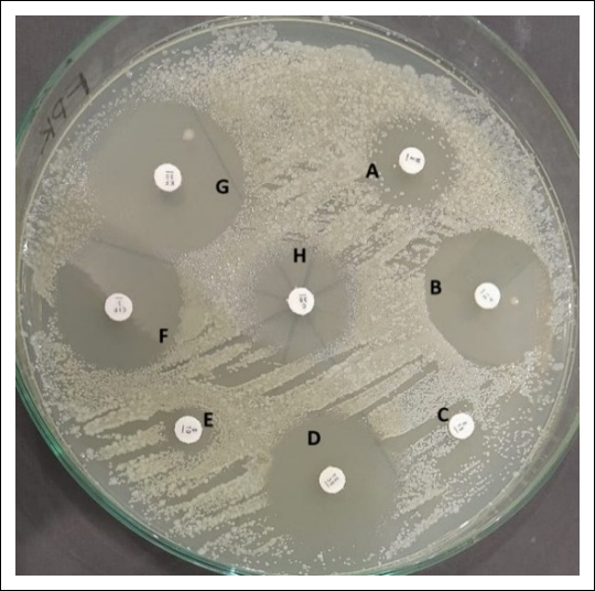
Figure 1. Representative antimicrobial susceptibility testing of presumptive Escherichia coli isolates using the Kirby–Bauer disk diffusion method. The image illustrates the inhibition zones produced by eight antimicrobial agents. Susceptibility categories were interpreted according to Clinical and Laboratory Standards Institute (CLSI) M100 (2024), CLSI M100-S25 for cephalothin, and National Committee for Clinical Laboratory Standards (NCCLS) M100-S11 for erythromycin. Labels indicate: (A) trimethoprim (25 mm, susceptible); (B) ampicillin (7 mm, resistant); (C) erythromycin (7.4 mm, resistant); (D) tetracycline (22.6 mm, susceptible); (E) streptomycin (8.6 mm, resistant); (F) ciprofloxacin (23.4 mm, susceptible); (G) cephalothin (12.4 mm, resistant); and (H) chloramphenicol (10.4 mm, resistant).
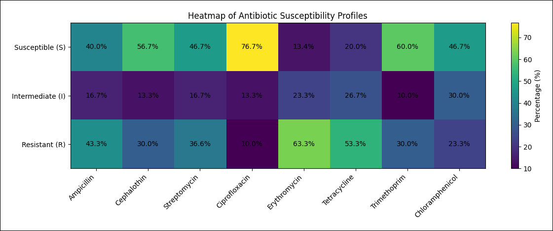
Figure 2. Heatmap of antimicrobial susceptibility profiles of Escherichia coli isolates recovered from raw goat milk samples.
Farm-wise distribution of MDR isolates
Farm-wise analysis demonstrated considerable variation in the occurrence of MDR isolates (Table 3). The highest proportion of MDR isolates was recorded in Farm A (50.0%), where three of the six E. coli isolates exhibited resistance to three or more antimicrobial classes. In contrast, no MDR isolate was detected in Farm C (0%). Overall, the prevalence of MDR among farms ranged from 0% to 50%.
The marked differences in MDR prevalence among farms indicate substantial heterogeneity in resistance patterns within the study area. The observed variation, ranging from 0% to 50%, provides novel farm-level information that has not previously been reported for goat-associated E. coli isolates in this region.
| Farm | Escherichia coli isolates (n) | Multidrug-resistant E. coli isolates (n) | Percentage |
|---|---|---|---|
| A | 6 | 3 | 50.0 |
| B | 4 | 1 | 25.0 |
| C | 1 | 0 | 0.0 |
| D | 5 | 2 | 40.0 |
| E | 7 | 2 | 28.6 |
| F | 3 | 1 | 33.3 |
| G | 4 | 1 | 25.0 |
| Total | 30 | 10 | 33.3 |
Table 3. Farm-wise distribution of multidrug-resistant E. coli isolates.
| Farm | Escherichia coli isolates (n) | Multidrug-resistant E. coli isolates (n) | Percentage |
|---|---|---|---|
| A | 6 | 3 | 50.0 |
| B | 4 | 1 | 25.0 |
| C | 1 | 0 | 0.0 |
| D | 5 | 2 | 40.0 |
| E | 7 | 2 | 28.6 |
| F | 3 | 1 | 33.3 |
| G | 4 | 1 | 25.0 |
| Total | 30 | 10 | 33.3 |
DISCUSSION
Occurrence of subclinical mastitis and isolation of E. coli
This study provides the first comprehensive phenotypic AMR profile of E. coli isolated from raw goat milk with subclinical mastitis in Siliragung Subdistrict, Banyuwangi, an important dairy goat production area characterized by widespread consumption of fresh goat milk. Although AMR in S. aureus has previously been documented in the same farms and β-lactamase genes, including TEM and SHV, have been detected in a limited number of E. coli isolates obtained from mixed goat and cattle samples in East Java, the present study provides previously unavailable district-specific disk diffusion data for eight antimicrobial agents. The findings revealed a distinct resistance pattern characterized by high resistance to erythromycin (63.3%) and tetracycline (53.3%), with 33.3% of isolates classified as MDR. These findings extend previous studies conducted in Indonesia, which generally involved fewer antimicrobial agents or focused on different geographic regions, and provide valuable information on the resistance characteristics of E. coli circulating in dairy goats.
A total of 91 of 224 milk samples (40.6%) were positive for subclinical mastitis based on CMT scores of 3 and 4. This prevalence is comparable to that reported by Ariffin et al. [26] in Malaysia and Öztürk et al. [27] in Turkey, both of whom reported prevoximately 40%. However, the prevalence observed in the present study was lower than that reported by Michira et al. [28] in Kenya (53%) and Al Emon et al. [29] in Bangladesh (64.92%). In Indonesia, Nuraini et al. [30] reported a higher national prevalence of mastitis in dairy animals (59.44%). Nevertheless, the prevalence observed in the present study was close to the regional prevalence of 44.96% reported by Praja et al. [22], who identified S. aureus as the causative agent of subclinical mastitis in Banyuwangi.
Despite the importance of E. coli as a major Gram-negative mastitis pathogen and foodborne zoonotic bacterium, information regarding its occurrence and resistance characteristics in Siliragung Subdistrict has been lacking. Although a recent study conducted in East Java detected β-lactamase genes in a limited number of goat milk-derived E. coli isolates, including several from Banyuwangi, that study did not provide detailed antimicrobial susceptibility profiles, evaluate farm-level variation, or focus exclusively on subclinical mastitis cases in goats. Therefore, the present study fills an important knowledge gap by providing district-specific phenotypic data and farm-level information.
Several factors may influence the occurrence of subclinical mastitis, including body condition score, stage of lactation, breed, and teat morphology [31]. The increased prevalence observed in older animals may be attributed to prolonged exposure to environmental pathogens compared with younger animals [32]. In addition, inadequate implementation of biosecurity measures may have contributed to the relatively high prevalence of subclinical mastitis observed in the present study. Such conditions favor the persistence and transmission of mastitis-causing bacteria, particularly S. aureus, which has been reported as the predominant pathogen in several previous studies conducted in Bangladesh, Kenya, and Malaysia [26, 28, 29].
In the present study, 30 of 91 CMT-positive samples (32.9%) were confirmed as E. coli. This isolation rate was markedly higher than that reported by Ariffin et al. [26] in Malaysia (1.1%) and Michira et al. [28] in Kenya (1.0%). Environmental E. coli mastitis is strongly influenced by management factors, including hygiene during milking and post-milking practices, which may facilitate pathogen invasion and colonization [34]. The relatively low occurrence of E. coli associated subclinical mastitis in the present study may reflect improvements in environmental sanitation and farm management practices within the study area. Nevertheless, the presence of E. coli in farms with low subclinical mastitis prevalence indicates that environmental contamination remains an important source of infection [33].
Antimicrobial susceptibility profiles of E. coli
The isolates in this study were tested against eight antimicrobial agents, namely ampicillin, cephalothin, streptomycin, ciprofloxacin, erythromycin, chloramphenicol, trimethoprim, and tetracycline (Table 2). Resistance was observed against several antimicrobial agents, whereas the highest susceptibility rates were recorded for ciprofloxacin and trimethoprim. Similar susceptibility to ciprofloxacin was reported by Sobur et al. [35]. However, unlike the findings of Sobur et al. [35], which reported tetracycline resistance, the isolates in the present study exhibited greater susceptibility to this antimicrobial agent.
The high susceptibility to ciprofloxacin may be explained by its mechanism of action, which targets two essential enzymes, namely DNA gyrase and topoisomerase IV. DNA gyrase represents the principal target in E. coli, and inhibition of this enzyme interferes with DNA supercoiling and replication [36]. Resistance to fluoroquinolones may arise through several mechanisms, including mutations in genes encoding DNA gyrase, increased drug efflux, and target protection mechanisms [37].
The relatively high susceptibility to trimethoprim observed in this study may reflect the limited use of this antimicrobial agent in the study area. Trimethoprim exerts its antibacterial activity by inhibiting dihydrofolate reductase [38]. Resistance to trimethoprim has been associated with the presence of the dfr gene [39]. Therefore, the susceptibility observed in the present study suggests that the isolates may not harbor genes responsible for trimethoprim resistance.
Tetracyclines are among the antimicrobial agents commonly used by farmers in Siliragung Subdistrict. Resistance to tetracyclines is generally associated with genes encoding tetracycline resistance determinants, including tetA-Y [40]. The susceptibility observed among several isolates in the present study may indicate the absence of these resistance determinants, although molecular studies are required to confirm this hypothesis.
Resistance to ampicillin, cephalothin, streptomycin, erythromycin, and chloramphenicol was also observed. Notably, erythromycin resistance was particularly high and was consistent with findings reported by Kallau et al. [41] and Sobur et al. [35]. In contrast, Ariffin et al. [26] and Balemi et al. [42] reported chloramphenicol susceptibility, whereas the chloramphenicol resistance observed in the present study was more consistent with the findings of Michira et al. [28]. The elevated resistance rates observed in the present study may be attributable to inappropriate and excessive use of antimicrobial agents [43].
The high prevalence of erythromycin resistance (63.3%) is noteworthy and may indicate an emerging resistance pattern in the study area. Empirical and uncontrolled antimicrobial usage among smallholder farmers, combined with limited awareness regarding AMR and inadequate regulatory oversight, may have contributed to this phenomenon. However, quantitative data on antimicrobial consumption were unavailable to support this assumption. Furthermore, the interpretation of erythromycin susceptibility in E. coli should be approached with caution because standardized breakpoints for macrolides in Gram-negative bacteria are limited and often extrapolated. Therefore, the findings of the present study highlight the importance of continued surveillance and further investigations.
High resistance to erythromycin may be mediated by erythromycin ribosomal methylation mechanisms associated with the erm gene, which interfere with the binding of erythromycin to the 50S ribosomal subunit [44]. The predominance of erythromycin resistance observed in the present study provides novel insight, as macrolide resistance has been reported inconsistently in environmental E. coli isolates associated with goat mastitis and is rarely the dominant resistance phenotype in comparable studies conducted in Southeast Asia.
Mechanisms of AMR and occurrence of MDR
Resistance of E. coli to β-lactam antibiotics is associated with alterations in the bacterial outer membrane that affect antimicrobial uptake, particularly changes in porin expression profiles [45]. Mutations in genes encoding outer membrane proteins, including OmpD, may reduce membrane permeability and contribute to resistance against β-lactam antibiotics. Furthermore, alterations in proteins involved in cell wall synthesis can influence susceptibility to ampicillin, which exerts its bactericidal activity by inhibiting bacterial cell wall synthesis during active replication [46]. In addition, bacteria may resist β-lactam antibiotics through the production of β-lactamases, modification of target proteins, reduced membrane permeability, and increased expression of drug efflux pumps [47].
Resistance to streptomycin is primarily associated with impaired transport of the antimicrobial agent into bacterial cells [48]. Moreover, the presence of genes encoding aminoglycoside adenyltransferases, such as aadA1, has been implicated in streptomycin resistance [49]. Similarly, resistance to chloramphenicol is frequently mediated by the production of chloramphenicol acetyltransferase enzymes, which inactivate the drug. Additional mechanisms, including target modification, drug efflux systems, and changes in outer membrane permeability, have also been reported [50].
MDR generally refers to resistance to more than two antimicrobial classes. One of the most widely accepted approaches for classifying bacteria as MDR is based on the results of in vitro antimicrobial susceptibility testing against multiple antimicrobial classes [51]. In the present study, E. coli isolates exhibited resistance to four antimicrobial classes, namely macrolides, aminoglycosides, phenicols, and β-lactams. The relatively high prevalence of MDR isolates observed in this study may be associated with the widespread use of broad-spectrum antimicrobial agents and antibiotic combinations in both animals and humans in the region [52].
Although AMR in E. coli has previously been reported in livestock, the present study differs from earlier investigations in several important aspects. Previous work by Praja et al. [10] focused on milk from dairy cattle collected in Licin Subdistrict, a highland area with lower ambient temperatures, whereas the present study investigated milk from dairy goats in Siliragung Subdistrict, a lowland region with a warmer climate. Such environmental differences may influence management practices and bacterial ecology. In addition, Dewi et al. [21] investigated fecal samples collected from livestock in a different geographical setting rather than milk samples. Therefore, differences in host species, sample type, and environmental conditions distinguish the present study from previous reports. To the best of our knowledge, this study represents the first report describing AMR in E. coli isolated from raw goat milk associated with subclinical mastitis in Siliragung Subdistrict, Banyuwangi, East Java, Indonesia.
MDR bacteria can acquire resistance through horizontal gene transfer and spontaneous mutations resulting from prolonged exposure to antimicrobial agents. Although the development of resistance is a natural evolutionary process, its emergence and dissemination are accelerated by inappropriate antimicrobial use, inadequate surveillance, and insufficient regulatory control [1]. Consequently, appropriate antimicrobial usage, improved biosecurity measures, increased awareness among livestock workers, and national surveillance programs are essential strategies for limiting the spread of resistant bacteria [53].
The primary objective of the present study was to investigate the occurrence of bacterial pathogens associated with subclinical mastitis in raw goat milk and to evaluate their antimicrobial susceptibility profiles. Accordingly, the study was intentionally designed to focus on phenotypic identification and susceptibility testing as an initial approach for characterizing the isolates. Molecular analyses were not included; however, they would provide valuable complementary information and could strengthen the present findings by confirming the presence of resistance genes in MDR E. coli isolates.
Strengths, limitations, and future perspectives
Despite the important findings, several limitations should be acknowledged. First, sampling was conducted during a limited period (June–December 2024), which may not fully capture seasonal variations in the occurrence of subclinical mastitis and AMR patterns. Second, the number of isolates obtained from individual farms was relatively small, potentially limiting the generalizability of the findings. Third, only milk samples with CMT scores of 3 and 4 were included, potentially introducing sampling bias. Finally, the absence of somatic cell count measurements alongside CMT results may have affected the accuracy of subclinical mastitis diagnosis. These limitations should be considered when interpreting the results, and future studies should incorporate larger sample sizes, extended sampling periods, and complementary diagnostic methods.
Notwithstanding these limitations, several strengths of the present study should be emphasized. This investigation represents the first district-specific phenotypic characterization of AMR in E. coli isolated from raw goat milk associated with subclinical mastitis in Siliragung Subdistrict. Furthermore, the inclusion of all seven active dairy goat farms and the evaluation of susceptibility against eight clinically relevant antimicrobial agents provided comprehensive farm-level information that has not previously been available for this production system.
New strategies are urgently needed to address the growing global burden of AMR [54]. A One Health approach should include enhanced public awareness, improved hygiene practices, prevention of the transmission of infections, strengthened surveillance of AMR and antimicrobial consumption in both humans and animals, development of rapid diagnostic tools, and promotion of vaccines and alternative therapeutic approaches [55]. In addition, the development of novel antimicrobial agents is both time-consuming and expensive. Consequently, repurposing approved drugs with demonstrated antibacterial activity represents a promising alternative. Such drugs may exhibit enhanced efficacy when used in combination therapies, provided that clinically safe and effective concentrations can be established. Because structural, pharmacological, bioavailability, safety, toxicity, and pharmacokinetic information is already available for these compounds, several stages of drug development can potentially be accelerated or bypassed [56].
By focusing exclusively on goat milk obtained from a well-defined smallholder production system and relating the findings to local consumption practices, the present study provides novel evidence to support mastitis control programs and One Health policies aimed at combating AMR in Indonesian dairy goat farming systems.
CONCLUSION
This study provides the first district-specific phenotypic characterization of AMR in E. coli isolated from raw goat milk associated with subclinical mastitis in dairy goats from Siliragung Subdistrict, Banyuwangi, Indonesia. Among 224 milk samples collected from seven dairy goat farms, 91 samples (40.6%) were positive for subclinical mastitis, and 30 isolates (32.9%) were confirmed as E. coli. The highest resistance rates were observed for erythromycin (63.3%) and tetracycline (53.3%), whereas the highest susceptibilities were recorded for ciprofloxacin (76.7%) and trimethoprim (60.0%). In addition, one-third of the isolates (33.3%) were classified as MDR, with marked variation in MDR prevalence among farms ranging from 0% to 50%.
These findings highlight the potential public health risks associated with consuming raw goat milk and emphasize the importance of prudent antimicrobial use, improved farm hygiene, enhanced biosecurity measures, and routine antimicrobial susceptibility testing. The farm-level variation observed in resistance patterns underscores the need for targeted interventions and surveillance programs tailored to smallholder dairy goat production systems. The study also provides valuable baseline information for veterinarians, livestock producers, and policymakers to support evidence-based antimicrobial stewardship and One Health strategies to limit the dissemination of resistant bacteria.
Overall, the present study provides novel and clinically relevant evidence regarding the occurrence and resistance patterns of E. coli associated with subclinical mastitis in dairy goats and contributes important baseline data for the development of effective mastitis control programs and One Health policies aimed at mitigating the growing threat of AMR in Indonesia.
DATA AVAILABILITY
The supplementary data can be made available from the corresponding author upon request.
GENERATIVE AI DECLARATION
The authors declare that generative Artificial Intelligence tools were used solely to improve language, grammar, and readability during manuscript preparation. All scientific content, data analysis, interpretation of results, and conclusions were developed and verified by the authors. The authors take full responsibility for the accuracy, integrity, and originality of the work presented, and no AI tool was listed as an author.
AUTHORS’ CONTRIBUTIONS
RNP: Conceptualization, methodology, supervision, and writing – review and editing. AY: Formal analysis, methodology, and writing – review and editing. JMH: Data curation, formal analysis, and writing – original draft. ALS: Data curation and sample processing. AB: Data curation, validation, and writing – review and editing. SW and AME: Data curation, methodology, and validation. HM and SSP: Laboratory samples, investigation, and inventory management. All authors have read and approved the final version of the manuscript.
COMPETING INTERESTS
The authors declare that they have no competing interests.
PUBLISHER’S NOTE
Veterinary World remains neutral with regard to jurisdictional claims in the published institutional affiliations.
ACKNOWLEDGMENTS
This research was funded by the Lembaga Penelitian dan Pengabdian Masyarakat (LPPM), Universitas Airlangga, Indonesia, through the project of Airlangga Research Fund 2024 (Grant Number: 672/UN3/2024). The authors gratefully acknowledge the Faculty of Health, Medicine, and Life Sciences (FIKKIA), Universitas Airlangga Banyuwangi Campus, for providing the laboratory facilities necessary for this study. The authors also extend their appreciation to the Veterinary Microbiology Research Team for their assistance with field sample collection and contributions to data analysis.
REFERENCES
- Salam MA, Al-Amin MY, Salam MT, Pawar JS, Akhter N, Rabaan AA. Antimicrobial resistance: a growing serious threat for global public health. Healthcare (Basel) 2023;11(13):1946. [Google Scholar]
- Sihombing B, Bhatia R, Srivastava R, Aditama TY, Laxminarayan R, Rijal S. Response to antimicrobial resistance in South-East Asia Region. Lancet Reg Health Southeast Asia 2023;18:100306. [Google Scholar]
- Xu T, Cao W, Huang Y, Zhao J, Wu X, Yang Z. The prevalence of Escherichia coli derived from bovine clinical mastitis and distribution of resistance to antimicrobials in part of Jiangsu Province, China. Agriculture 2023;13(1):90. [Google Scholar]
- Wilm J, Svennesen L, Kirkeby C, Krömker V. Treatment of clinically severe bovine mastitis – a scoping review. Front Vet Sci 2024;11:1286461. [Google Scholar]
- Lestari DS, Setiawan B, Saputro AL, Mafruchati M, Praja RN, Prastiya RA. Environmental and management risk factors for goat mastitis in Banyuwangi. JBMV 2025;14(2):202-212. [Google Scholar]
- Khasapane NG, de Smidt O, Lekota KE, Nkhebenyane J, Thekisoe O, Ramatla T. Antimicrobial resistance and virulence determinants of Escherichia coli isolates from raw milk of dairy cows with subclinical mastitis. Animals 2025;15(13):1980. [Google Scholar]
- Naidoo N, Zishiri OT. Presence, pathogenicity, antibiotic resistance, and virulence factors of Escherichia coli: a review. Bacteria 2025;4(1):16. [Google Scholar]
- Sarba EJ, Wirtu W, Gebremedhin EZ, Borena BM, Marami LM. Occurrence and antimicrobial susceptibility patterns of Escherichia coli and Escherichia coli O157 isolated from cow milk and milk products, Ethiopia. Sci Rep 2023;13:16018. [Google Scholar]
- Checcucci A, Buscaroli E, Modesto M, Luise D, Blasioli S, Scarafile D. The swine waste resistome: spreading and transfer of antibiotic resistance genes in Escherichia coli strains and the associated microbial communities. Ecotoxicol Environ Saf 2024;283:116774. [Google Scholar]
- Praja RN, Yudhana A, Pratama LD, Pratiwi EK, Herina RB, Setyarini DW. Antibiotic resistance of Escherichia coli isolated from dairy cow milk associated with subclinical mastitis in the Licin Subdistrict, East Java, Indonesia. Open Vet J 2025;15(11):6009-6019. [Google Scholar]
- Kadri SS. Key takeaways from the U.S. CDC's 2019;48(7):939-945. [Google Scholar]
- Karwowska E. Antibiotic resistance in the farming environment. Appl Sci 2024;14(13):5776. [Google Scholar]
- Belay WY, Getachew M, Tegegne BA, Teffera ZH, Dagne A, Zeleke TK. Mechanism of antibacterial resistance, strategies and next-generation antimicrobials to contain antimicrobial resistance: a review. Front Pharmacol 2024;15:1444781. [Google Scholar]
- Galgano M, Pellegrini F, Catalano E, Capozzi L, Del Sambro L, Sposato A. Acquired bacterial resistance to antibiotics and resistance genes: from past to future. Antibiotics 2025;14(3):222. [Google Scholar]
- Floris I, Battistini R, Tramuta C, Garcia-Vozmediano A, Musolino N, Scardino G. Antibiotic resistance in lactic acid bacteria from dairy products in Northern Italy. Antibiotics 2025;14(4):375. [Google Scholar]
- Sanjulián L, Fernández-Rico S, González-Rodríguez N, Cepeda A, Miranda JM, Fente C. The role of dairy in human nutrition: myths and realities. Nutrients 2025;17(4):646. [Google Scholar]
- Onoharigho FO, Ighede PA, Edo GI, Akpoghelie PO, Akpoghelie EO. Isolation and identification of bacterial and fungal spoilage organisms in branded and unbranded milk; consumer perception of safety hazard for milk. Appl Microbiol Theory Technol 2022;3:31-48. [Google Scholar]
- Al-Kaisy QH, Al-Saadi JS, Al-Rikabi AKJ, Altemimi AB, Hesarinejad MA, Abedelmaksoud TG. Exploring the health benefits and functional properties of goat milk proteins. Food Sci Nutr 2023;11(10):5641-5656. [Google Scholar]
- Zhu L, Fan Z, Li W, Shan Y. Goat milk exhibits a higher degree of protein oxidation and aggregation than cow milk during cold storage. Foods 2025;14(5):852. [Google Scholar]
- Liao G, Wang T, Li X, Gu J, Jia Q, Wang Z. Comparison of the lipid composition of milk fat globules in goat (Capra hircus) milk during different lactations and human milk. Foods 2024;13(11):1618. [Google Scholar]
- Dewi RR, Nuryawan A, Jajere SM, Sihombing JM, Tambunan IJ. Antimicrobial resistance profiles of Escherichia coli derived from an integrated agroforestry-livestock system in Deli Serdang Regency, Indonesia. Vet World 2024;17(3):690-699. [Google Scholar]
- Praja RN, Yudhana A, Saputro AL, Hamonangan JM. The first study on antimicrobial resistance of Staphylococcus aureus isolated from raw goat milk associated with subclinical mastitis in Siliragung Subdistrict, East Java, Indonesia. Vet World 2023;16:786-791. [Google Scholar]
- Faizah AN, Setiawan B, Saputro AL, Warsito SH, Praja RN, Fikri F. Isolation, identification and risk factors of Staphylococcus aureus bacteria in dairy goat milk with subclinical mastitis in Siliragung District, Banyuwangi Regency. JBMV 2023;12(2):68-72. [Google Scholar]
- Nanda ERV, Harijani N, Wibawati PA. Uji total bakteri susu segar kambing Jawa Randu di Siliragung, Banyuwangi. J Med Vet 2020;3(2):224-229. [Google Scholar]
- Magiorakos AP, Srinivasan A, Carey RB, Carmeli Y, Falagas ME, Giske CG. Multidrug-resistant, extensively drug-resistant and pandrug-resistant bacteria: an international expert proposal for interim standard definitions for acquired resistance. Clin Microbiol Infect 2012;18(3):268-281. [Google Scholar]
- Ariffin S, Hasmadi N, Syawari N, Sukiman M, Ariffin M, Chai M. Prevalence and antibiotic susceptibility pattern of Staphylococcus aureus, Streptococcus agalactiae and Escherichia coli in dairy goats with clinical and subclinical mastitis. J Anim Health Prod 2019;7:32-37. [Google Scholar]
- Öztürk D, Türütoğlu H, Pehlivanoğlu F, Yapıcıer ÖŞ. Identification of bacteria isolated from dairy goats with subclinical mastitis and investigation of methicillin and vancomycin resistant Staphylococcus aureus strains. Ankara Üniv Vet Fak Derg 2019;66:191-196. [Google Scholar]
- Michira L, Kagira J, Maina N, Waititu K, Kiboi D, Ongera E. Prevalence of subclinical mastitis, associated risk factors and antimicrobial susceptibility pattern of bacteria isolated from milk of dairy cattle in Kajiado Central sub-county, Kenya. Vet Med Sci 2023;9(6):2885-2892. [Google Scholar]
- Al Emon A, Hossain H, Chowdhury MSR, Rahman MA, Tanni FY, Asha MN. Prevalence, antimicrobial susceptibility profiles and resistant gene identification of bovine subclinical mastitis pathogens in Bangladesh. Heliyon 2024;10(14):e34567. [Google Scholar]
- Nuraini DM, Andityas M, Sukon P, Phuektes P. Prevalence of mastitis in dairy animals in Indonesia: a systematic review and meta-analysis. Vet World 2023;16:1380-1389. [Google Scholar]
- Algharib SA, Dawood AS, Huang L, Guo A, Zhao G, Zhou K. Basic concepts, recent advances, and future perspectives in the diagnosis of bovine mastitis. J Vet Sci 2024;25(1):e18. [Google Scholar]
- Khasapane NG, Byaruhanga C, Thekisoe O, Nkhebenyane SJ, Khumalo ZTH. Prevalence of subclinical mastitis, its associated bacterial isolates and risk factors among cattle in Africa: a systematic review and meta-analysis. BMC Vet Res 2023;19(1):123. [Google Scholar]
- Lima MC, Souza MC, Espeschit IF, Maciel PA, Sousa JE, Moraes GF. Mastitis in dairy goats from the state of Minas Gerais, Brazil: profiles of farms, risk factors and characterization of bacteria. Pesq Vet Bras 2018;38:1742-1751. [Google Scholar]
- Zaatout N. An overview on mastitis-associated Escherichia coli: pathogenicity, host immunity and the use of alternative therapies. Microb Res 2022;256:126960. [Google Scholar]
- Sobur MA, Sabuj AAM, Sarker R, Rahman AT, Kabir SL, Rahman MT. Antibiotic-resistant Escherichia coli and Salmonella spp. associated with dairy cattle and farm environment having public health significance. Vet World 2019;12:984. [Google Scholar]
- Shariati A, Arshadi M, Khosrojerdi MA, Abedinzadeh M, Ganjalishahi M, Maleki A. The resistance mechanisms of bacteria against ciprofloxacin and new approaches for enhancing the efficacy of this antibiotic. Front Public Health 2022;10:1025633. [Google Scholar]
- El-sagheir AMK, Wenzel M, Yli-Kauhaluoma J. Fluoroquinolones as versatile scaffolds: potential for targeting classical and novel mechanisms to combat antibacterial resistance. Eur J Pharm Sci 2025;214:107247. [Google Scholar]
- Tjampakasari CR. Dihydrofolate reductase (dfr) and dihydropteroate synthase (sul) gene mutations in Escherichia coli trimethoprim-sulfamethoxazole resistance. Indonesian J Biotechnol Biodivers 2024;8(1):51-59. [Google Scholar]
- Spencer A, Wong Q, Lawson ST, Fry H, Ramchandani NM, Harding C. Trimethoprim resistance in Escherichia coli exhibits an allele-specific growth advantage. J Med Microbiol 2025;74(6):002021. [Google Scholar]
- Zhao X, Zhang M, Zhang Z, Wang L, Wang Y, Liu L. Guanethidine restores tetracycline sensitivity in multidrug-resistant Escherichia coli carrying tetA gene. Antibiotics 2024;13(10):973. [Google Scholar]
- Kallau NHG, Wibawan IWT, Lukman DW, Sudarwanto MB. Detection of multi-drug resistant (MDR) Escherichia coli and tet gene prevalence at a pig farm in Kupang, Indonesia. J Adv Vet Anim Res 2018;5:388. [Google Scholar]
- Balemi A, Gumi B, Amenu K, Girma S, Gebru MU, Tekle M. Prevalence of mastitis and antibiotic resistance of bacterial isolates from CMT positive milk samples obtained from dairy cows, camels, and goats in two pastoral districts in Southern Ethiopia. Animals 2021;11:1530. [Google Scholar]
- Ahmed SK, Hussein S, Qurbani K, Ibrahim RH, Fareeq A, Mahmood KA. Antimicrobial resistance: impacts, challenges, and future prospects. J Med Surg Public Health 2024;2:100081. [Google Scholar]
- Amdan NAN, Shahrulzamri NA, Hashim R, Jamil NM. Understanding the evolution of macrolides resistance: a mini review. J Glob Antimicrob Resist 2024;38:368-375. [Google Scholar]
- Saini J, Gautam S, Sharma D, Khanduri A, Venugopal D. Revisiting the metallo-β-lactamase-mediated antibiotic resistance: exploring novel mechanisms and therapeutic strategies. Int J Microbiol 2025;2025:1574819. [Google Scholar]
- Türkyılmaz O, Darcan C. Molecular basis of ampicillin resistance: combinatorial mechanisms and future strategies. World J Microbiol Biotechnol 2026;42(2):79. [Google Scholar]
- Li M, Liu Q, Teng Y, Ou L, Xi Y, Chen S. The resistance mechanism of Escherichia coli induced by ampicillin in laboratory. Infect Drug Resist 2019;12:2853-2863. [Google Scholar]
- Lang M, Carvalho A, Baharoglu Z, Mazel D. Aminoglycoside uptake, stress, and potentiation in Gram-negative bacteria: new therapies with old molecules. Microbiol Mol Biol Rev 2023;87(4):e0003622. [Google Scholar]
- Stasiak M, Maćkiw E, Kowalska J, Kucharek K, Postupolski J. Silent genes: antimicrobial resistance and antibiotic production. Pol J Microbiol 2021;70(4):421-429. [Google Scholar]
- Jaiswal A, Khan A, Yogi A, Singh S, Pal AK, Soni R. Isolation and molecular characterization of multidrug resistant Escherichia coli from chicken meat. 3 Biotech 2024;14(4):107. [Google Scholar]
- Jaglal P, Velaphi SC, Menezes CN, Swe Swe-Han K. In vitro evaluation of antimicrobial synergy against multidrug-resistant Gram-negative paediatric bloodstream pathogens in South Africa. Antibiotics 2025;14(7):630. [Google Scholar]
- Matheou A, Abousetta A, Pascoe AP, Papakostopoulos D, Charalambous L, Panagi S. Antibiotic use in livestock farming: a driver of multidrug resistance? Microorganisms 2025;13(4):779. [Google Scholar]
- Eldesoukey IE, Elmonir W, Alouffi A, Beleta EI, Kelany MA, Elnahriry SS. Multidrug-resistant enteropathogenic Escherichia coli isolated from diarrhoeic calves, milk, and workers in dairy farms: a potential public health risk. Antibiotics 2022;11:999. [Google Scholar]
- Patra M, Gupta AK, Kumar D, Kumar B. Antimicrobial resistance: a rising global threat to public health. Infect Drug Resist 2025;18:5419-5437. [Google Scholar]
- Al-Khalaifah H, Rahman MH, Al-Surrayai T, Al-Dhumair A, Al-Hasan M. A One-Health perspective of antimicrobial resistance (AMR): human, animals and environmental health. Life 2025;15(10):1598. [Google Scholar]
- Panchal YS, Patel AS, Vadgama HA, Parmar GS, Joshi BA. Drug repurposing: new antimicrobial applications of non-antibiotic drugs in veterinary medicine. Int J Adv Res 2025;13(12):934-944. [Google Scholar]